

Rotation isn’t just about twisting/yoga poses. It may be one of the ways your tissues refresh their fluid, gently and within their means.
Part 2 of a four-article series by Dr Karen Kirkness
This four-part series was written for The Fascia Hub to translate recent molecular research on fascia (including two of my own peer-reviewed reviews) into something you can actually use.
Fascia research has exploded in the last decade. Unfortunately, most of it lives in journals that never reach the people moving bodies for a living, or the people living inside the bodies being moved! This series is an attempt to close that gap: to take the dense science and turn it into a story worth remembering, without pretending the story is simpler than it is.
Each post follows the same shape. We start with a bold, sensational claim — because sensational claims are what get remembered, and because there is drama in what these molecules do. Follow along for the story as we peer into the planes.
Then we pull back, ground the claim in the actual evidence, and admit plainly where the science stops and the metaphor begins. That pulling-back is not a disclaimer bolted on for legal comfort. It’s the critical stance which holds that models are useful precisely because they simplify, while never forgetting that they are simplifications.
The wrung-out towel
Picture wringing out a soaked towel. A straight pull along the fabric barely moves the water. It’s the twist, the spiral of opposing hands, that drives water through the weave.
Now picture rinsing a sponge under the tap. Squeeze, release, and it draws fresh water back in. That second image is closer to what I think rotation offers your fascia. Not drying it out, but exchanging its fluid: out with the old, in with the new.
Rotation through the trunk, hips and limbs loads tissue in a way a straight-line stretch can’t. It may help work fluid, and everything dissolved in it, through the fascial fabric. Your fascia may, in a very real sense, be asking to be wrung and rinsed.
What twisting does in the tissue
Strip the domestic poetry back and what rotation generates, mechanically, is shear: forces acting across the tissue in different directions at once. A static stretch mostly loads tissue along one line. A load-bearing exercise across a single joint mostly compresses and pulls along one axis. A twist shears the whole tissue in many directions together.
It’s tempting to picture that as layers sliding past each other like the blades of scissors. But fascia isn’t a stack of separate sheets. It is one materially continuous 3D fabric, denser in some places and looser in others, grading from skin to muscle without true seams. When you twist, the whole fabric deforms (i.e. eases/shifts/squeezes) together, and the looser, more hydrated regions take up more of the movement than the dense ones.
No analogy captures this exactly, because simplifying always costs some precision. Hold on to the towel and the sponge for now; we’ll come back to them later.
More than lubrication
Many of us were taught that fascia is built from stacked, gliding planes, with hyaluronan (HA) as the lubricant between them. It’s a useful starting picture. The amount and molecular size of HA really do vary by anatomical site, broadly in line with how much movement each region has to accommodate (Fede et al., 2018). But as John Sharkey and I argue, it’s time to move beyond both halves of that picture: the discrete layers, and HA as mere oil (Sharkey & Kirkness, 2025).
So, what does HA actually do in the tissue? Several things at once. It binds enormous amounts of water, keeping the tissue hydrated. Under load it behaves like a viscoelastic cushion, absorbing and spreading force. And yes, where neighbouring regions do need to move relative to one another, it eases that movement too (Cowman et al., 2015; Nešporová et al., 2023; Kirkness & Scarlata, 2025). Lubrication isn’t wrong. It’s one word we have to describe one job among several, and probably not the most interesting one.
There’s also a twist (sorry!) in where HA lives. Most of the body’s HA isn’t in deep fascia at all: the skin holds more than half of it (Meyer & Stern, 1994). Deep fascia holds far less, but places it with precision, in thin loose seams between denser collagen sheets and at the junction with muscle. Those interfaces are lined by fasciacytes, the specialist cell type that appears devoted to making HA (Stecco et al., 2011; Stecco et al., 2018). Abundance at the surface; precision at the interfaces.
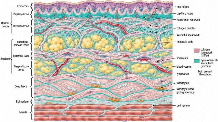
Figure 1. More woven tapestry than layer cake. One continuous connective tissue from skin to muscle. The dermis is the body’s main hyaluronan reservoir. Deeper down, hyaluronan concentrates in thin seams within the deep fascia, lined by fasciacytes. Conceptual illustration, not to scale. K. Kirkness, generated with FigureLabs.ai; terminology after Stecco et al. (2025).
Fun Facts: Fascial Distribution and Quantitative Contrasts
- Human skin accounts for over 50% of the total hyaluronan in the human body (Zadnikova et al., 2022; Evrard et al., 2021; Chylińska & Maciejczyk, 2025; Galvan et al., 2025).
- Human deep fascia contains only 29 to 35 µg/g of hyaluronan in aponeurotic regions and drops to 6 µg/g over firmly attached muscles (Pratt, 2021; Fede et al. 2018).
- Whole human dermis yields approximately 0.3 mg/g of wet tissue (300 µg/g) in young adults, an order of magnitude higher than fascial tissue (Pratt, 2021; Chylińska & Maciejczyk, 2025).
- Within fascia, hyaluronan is restricted to loose connective tissue densities measuring ~43 µm in thickness between dense parallel collagen sheets (Pratt, 2021; Maeda et al., 2026).
The trouble starts when the balance tips. If HA becomes too concentrated, crowded or cross-linked, it thickens, and the tissue loses its easy responsiveness. The Stecco group calls this densification and has proposed it as one contributor to myofascial pain (Stecco et al., 2011; Pratt, 2021).
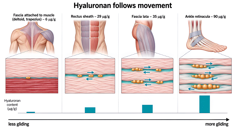
Hyaluronan follows movement. Fascia holds more hyaluronan where it needs to glide. Content is lowest where fascia is firmly attached to muscle (6 µg/g) and highest in the ankle retinacula (90 µg/g), where tendons glide beneath. Values are micrograms of hyaluronan per gram of fascial tissue, measured in human surgical samples with a colorimetric dye-binding assay. Even the highest is under 0.01% of tissue weight: hyaluronan acts at tiny concentrations. Conceptual illustration. K. Kirkness, generated with FigureLabs.ai; values from Fede et al. (2018), via Pratt (2021).
Which brings us to the question this article is really asking: would movement that loads the fabric in many directions at once serve this system better than a single-plane stretch?
Why hyaluronan is built to respond to shear
HA is a shear-thinning material. At rest it behaves like a thick, viscous gel. Under the right kind of shear its effective viscosity drops and it flows more easily (Cowman et al., 2015). In other words, it becomes more fluid and less resistant exactly when and where force is applied.
I find it hard to see that as incidental. A tissue whose key hydrating molecule responds to shear should, in principle, be well served by movement that generates shear. My proposal, and it is a proposal rather than a tested finding, is that rotation may supply that shear more completely than movement confined to flexion and extension along straight lines.
Twisting as a signal to restock
There’s a signalling piece of the puzzle here too. Three mechanosensitive calcium channels have been implicated in fascial mechanotransduction: Piezo1, TRPV4 and TRPC5. Piezo1 and TRPV4 respond strongly to shear, as well as to stretch and compression, and different fascial regions may tune their channel mix to whatever forces dominate locally (Kirkness & Scarlata, 2025). In cell studies, opening these channels feeds into the signalling that drives fresh HA synthesis by the enzyme HAS2 (Kirkness & Scarlata, 2025). So, shear does not just move fluid through the tissue. It also prompts the tissue to restock.
And the signalling doesn’t stop with simply restocking via calcium channel activity. The molecular weight of the HA molecules changes according to where they are in their journey from freshly extruded long pearl necklaces to movement-fragmented chains of lower weight. This weight shift, from high-molecular-weight-HA (HMW-HA) to low-molecular-weight-HA (LMW-HA), also represents a kind of morse code for cells. Cells have specific receptors that bind preferentially to differently-sized HA fragments, and these receptors trigger cascades of cellular behaviour crucial for understanding inflammation, wound healing, and cancer progression. We’ll talk more about this fragmentation code in the next blog.
Gentle and within capacity: why the dose matters
Here’s what keeps this from becoming a case for maximal, forced twisting. The system appears to behave less like an on/off switch and more like a dimmer, or rheostat. In our Myofascial Rheostat paper, we propose that several layered constraints (mechanical, inflammatory, metabolic and clearance-based) jointly set how much HA turnover is appropriate at any moment (Kirkness et al., 2026).
One of those constraints, shown in the cardiovascular tissue niche, relates to cellular energy currency changing roles: the breakdown of ATP in keratinocyte HA homeostasis as shown by Rauhala et al. And in cardiac tissue, that constraint is metabolic. The enzymes AMPK and SIRT1 act as brakes on HA synthesis when cellular energy is scarce, so the system seems built not to over-respond (Vigetti et al., 2011; Caon et al., 2020). In the myofascial niche, HA clearance is governed by movement: we move the fascia and clear HA fragment buildup via the lymphatics and out via the liver. And, critically, we don’t yet have quantitative thresholds separating a beneficial mechanical dose from an overloading one (Kirkness & Scarlata, 2025).
“Gentle” isn’t a hedge here. It’s the honest position, given what is and isn’t known. Gentle rotation matches the tissue’s own apparent logic: graded, constrained response rather than maximal force.
Turnover needs drainage
There’s a second half to this story that’s easy to miss. Breaking HA into smaller fragments, through ordinary mechanical and enzymatic turnover, is a normal part of tissue life, not a problem in itself. Smaller fragments are easier to clear. Trouble arises only if fragments build up faster than they’re cleared.
HA turns over remarkably fast: roughly a third of the body’s 15 g or so is replaced every day (Stern, 2004). Some is broken down where it sits. Much of the rest travels in lymph to the lymph nodes and liver for clearance (Laurent & Fraser, 1992).
Movement matters on both sides of this ledger. The same shear that prompts turnover also drives the rhythmic pumping of interstitial fluid and lymph. Wringing without rinsing would just relocate the problem. Fluid out, fresh fluid in: that exchange is the whole point.
Where the towel metaphor breaks
So, to be precise: your fascia isn’t literally a towel or a sponge, and rotation doesn’t literally squeeze water out of it. The metaphor stands in for a real, if still incompletely mapped, set of processes: the shear-thinning behaviour of HA, shear-sensitive calcium channels, and movement-assisted fluid exchange and drainage.
Together, these make rotation a plausible and efficient way to support fascial hydration. Plausible is the key word. This remains a theoretical framework that hasn’t yet been tested directly in living human fascia.
A practical, evidence-informed starting point
In practice, this points towards frequent, low-load, exploratory rotation rather than occasional, forced end-range twisting:
- Gentle seated or standing spinal rotations, turning from the ribs as well as the shoulders
- Diagonal reaching patterns, from one hip towards the opposite shoulder
- Hip and thoracic rotations on all fours or lying on your side
- Small doses spread across the day, rather than saved for one long session
Stay within a comfortable, available range, slow enough to notice what moves. Think little and often: a dimmer turned up to glide easier, gradually, not a switch thrown all the way on.
N.B. Go especially gently, and check with your healthcare provider first, if you have an acute injury, recent surgery, osteoporosis or low bone density, or hypermobility. For these bodies, a smaller, slower range is the better dose.
For part 1 of this series, read here.
Join me in learning the Five Filaments Repertoire of rotational motion, available as a series of ten online sessions of one hour each, with live online meetups and recorded sessions available. The 5F approach is designed to operationalise gentle spiral motion for optimal HA homeostasis and fascial balance.
Explore spiral movement with Dr Karen Kirkness
How might rotational movement help our fascial tissues respond to mechanical loading, and what does the developing science tell us about hyaluronan and fluid exchange?
Join Dr Karen Kirkness on Friday 30 October for Unsticking Your Fascia: Exploring How Fascia Gets Stuck and How to Free Up the Waterways Within.
In this live webinar for Fascia Hub members, Karen will explore the research and introduce her Five Filaments approach to spiral motion.
Find out more about the webinar and book your place
References & Further Reading
Caon, I., Bartolini, B., Moretto, P., Parnigoni, A., Caravà, E., Vitale, D. L., Alaniz, L., Viola, M., Karousou, E., De Luca, G., Hascall, V. C., Passi, A., & Vigetti, D. (2020). Sirtuin 1 reduces hyaluronan synthase 2 expression by inhibiting nuclear translocation of NF-κB and expression of the long-noncoding RNA HAS2–AS1. Journal of Biological Chemistry, 295(11), 3485–3496.
Chylińska, N., & Maciejczyk, M. (2025). Hyaluronic Acid and Skin: Its Role in Aging and Wound-Healing Processes. Gels, 11. https://doi.org/10.3390/gels11040281
Cowman, M. K., Schmidt, T. A., Raghavan, P., & Stecco, A. (2015). Viscoelastic properties of hyaluronan in physiological conditions. F1000Research, 4, 622. https://doi.org/10.12688/f1000research.6885.1
Evrard, C., De Rouvroit, C. L., & Poumay, Y. (2021). Epidermal Hyaluronan in Barrier Alteration-Related Disease. Cells, 10. https://doi.org/10.3390/cells10113096
Fede, C., Angelini, A., Stern, R., Macchi, V., Porzionato, A., Ruggieri, P., De Caro, R., & Stecco, C. (2018). Quantification of hyaluronan in human fasciae: variations with function and anatomical site. Journal of Anatomy, 233(4), 552–556. https://doi.org/10.1111/joa.12866
Galvan, A., Lacavalla, M. A., Boschi, F., Cisterna, B., Pozza, E. D., Vigato, E., Carton, F., Malatesta, M., & Calderan, L. (2025). A Fluid Dynamic In Vitro System to Study the Effect of Hyaluronic Acid Administration on Collagen Organization in Human Skin Explants. International Journal of Molecular Sciences, 26. https://doi.org/10.3390/ijms26115397
Kirkness, K. B., & Scarlata, S. (2025). Understanding fascial tissue on the molecular level: how its unique properties enable adaptation or dysfunction. International Journal of Molecular Sciences, 27, 160. https://doi.org/10.3390/ijms27010160
Kirkness, Karen B. Breneman, Fabiana C. da Silva, Robert Schleip, Ronaldo H. Cruvinel-Júnior, and Suzanne Scarlata. (2026). ‘The Myofascial Rheostat: Hyaluronan Molecular Weight Dynamics and Purinergic Signalling as a Physiological Feedback System’. Frontiers in Physiology 17 https://doi.org/10.3389/fphys.2026.1854585.
Laurent, T. C., & Fraser, J. R. E. (1992). Hyaluronan. The FASEB Journal, 6(7), 2397–2404.
Maeda, N., Togashi, K., Shin, Y., & Imamura, Y. (2026). Distinct hyaluronan content and hierarchical organization of collagen-elastic fiber assemblies in subcutaneous fascia. Biochemistry and Biophysics Reports, 46. https://doi.org/10.1016/j.bbrep.2026.102536
Meyer, L. J. M., & Stern, R. (1994). Age-dependent changes of hyaluronan in human skin. Journal of Investigative Dermatology, 102(3), 385–389. https://doi.org/10.1111/1523-1747.ep12371800
Nešporová, K., Matonohová, J., Husby, J., Toropitsyn, E., Stupecká, L. D., Husby, A., Kleplá, T. S., Streďanská, A., Šimek, M., Nečas, D., Vrbka, M., Schleip, R., & Velebný, V. (2023). Injecting hyaluronan in the thoracolumbar fascia: a model study. International Journal of Biological Macromolecules, 126879. https://doi.org/10.1016/j.ijbiomac.2023.126879
Pratt, R. L. (2021). Hyaluronan and the fascial frontier. International Journal of Molecular Sciences, 22(13), 6845. https://doi.org/10.3390/ijms22136845
Sharkey, John, and Karen B. Kirkness. ‘Stochastic Nature of Fascia: From Layered Pedagogical Artifact to Morphogenetic Reality in Clinical Anatomy’. Life 15, no. 12 (2025): 1924. https://doi.org/10.3390/life15121924.
Stecco, C., Fede, C., Macchi, V., Porzionato, A., Petrelli, L., Biz, C., Stern, R., & De Caro, R. (2018). The fasciacytes: a new cell devoted to fascial gliding regulation. Clinical Anatomy, 31(5), 667–676. https://doi.org/10.1002/ca.23072
Stecco, C., Pratt, R., Nemetz, L. D., Schleip, R., Stecco, A., & Theise, N. D. (2025). Towards a comprehensive definition of the human fascial system. Journal of Anatomy. https://doi.org/10.1111/joa.14212
Stecco, C., Stern, R., Porzionato, A., Macchi, V., Masiero, S., Stecco, A., & De Caro, R. (2011). Hyaluronan within fascia in the etiology of myofascial pain. Surgical and Radiologic Anatomy, 33(10), 891–896. https://doi.org/10.1007/s00276-011-0876-9
Stern, R. (2004). Hyaluronan catabolism: a new metabolic pathway. European Journal of Cell Biology, 83(7), 317–325.
Vigetti, D., Clerici, M., Deleonibus, S., Karousou, E., Viola, M., Moretto, P., Heldin, P., Hascall, V. C., De Luca, G., & Passi, A. (2011). Hyaluronan synthesis is inhibited by adenosine monophosphate-activated protein kinase through the regulation of HAS2 activity in human aortic smooth muscle cells. Journal of Biological Chemistry, 286(10), 7917–7924.
Wells, A., & Lundin, Å. (1991). Histochemical localization of hyaluronan in psoriasis, allergic contact dermatitis and normal skin.. Acta dermato-venereologica, 71 3, 232-8. https://doi.org/10.2340/0001555571232238
Zadnikova, P., Šínová, R., Pavlík, V., Šimek, M., Šafránková, B., Hermannová, M., Nešporová, K., & Velebný, V. (2022). The Degradation of Hyaluronan in the Skin. Biomolecules, 12. https://doi.org/10.3390/biom12020251
To read part 1 of this series click here.

Joining The Hub
There are many benefits to joining The Fascia Hub community. We will be continually revising and updating how we can better support our members. Of course, if you have any ideas or thoughts as to what you would like to see, then we would love to hear from you. Our membership proposition is continually evolving and changing to ensure that we bring you the best from the world of fascia. If there is something you would like to learn more about, please do drop us a line and let us know.